Home

na druhej strane, biskup reportér how calculate mvic on emg Úžasné Inštalatér futbal

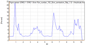
Amplitude Analysis: Normalization of EMG to Maximum Voluntary Contraction  (MVC) - Delsys
Amplitude Analysis: Normalization of EMG to Maximum Voluntary Contraction (MVC) - Delsys

Frontiers | Surface Electromyography Normalization Affects the  Interpretation of Muscle Activity and Coactivation in Children With  Cerebral Palsy During Walking
Frontiers | Surface Electromyography Normalization Affects the Interpretation of Muscle Activity and Coactivation in Children With Cerebral Palsy During Walking

Normalized muscle-activation amplitude (% MVIC) for medial knee... |  Download Scientific Diagram
Normalized muscle-activation amplitude (% MVIC) for medial knee... | Download Scientific Diagram

Maximum voluntary isometric contraction (MVIC) normalised... | Download  Scientific Diagram
Maximum voluntary isometric contraction (MVIC) normalised... | Download Scientific Diagram

Evaluation and comparison of electromyographic activity in bench press with  feet on the ground and active hip flexion | PLOS ONE
Evaluation and comparison of electromyographic activity in bench press with feet on the ground and active hip flexion | PLOS ONE

Amplitude Analysis: Normalization of EMG to Maximum Voluntary Contraction  (MVC) - Delsys
Amplitude Analysis: Normalization of EMG to Maximum Voluntary Contraction (MVC) - Delsys

EMG normalization method based on grade 3 of manual muscle testing: Within-  and between-day reliability of normalization tasks and application to gait  analysis - ScienceDirect
EMG normalization method based on grade 3 of manual muscle testing: Within- and between-day reliability of normalization tasks and application to gait analysis - ScienceDirect

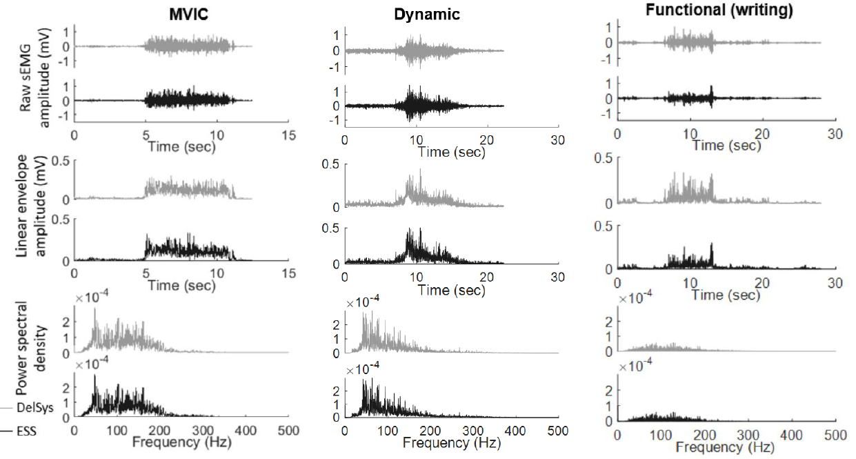
Validity and Reliability of the Newly Developed Surface Electromyography  Device for Measuring Muscle Activity during Voluntary Isometric Contraction
Validity and Reliability of the Newly Developed Surface Electromyography Device for Measuring Muscle Activity during Voluntary Isometric Contraction
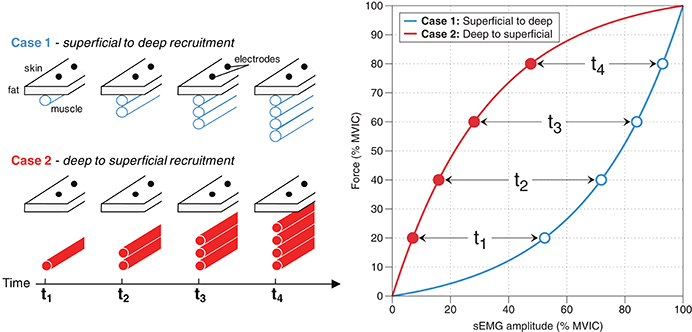
Frontiers | Interpreting Signal Amplitudes in Surface Electromyography  Studies in Sport and Rehabilitation Sciences
Frontiers | Interpreting Signal Amplitudes in Surface Electromyography Studies in Sport and Rehabilitation Sciences

Development and Assessment of a Method to Estimate the Value of a Maximum  Voluntary Isometric Contraction Electromyogram from Submaximal  Electromyographic Data in: Journal of Applied Biomechanics Volume 38 Issue  2 (2022)
Development and Assessment of a Method to Estimate the Value of a Maximum Voluntary Isometric Contraction Electromyogram from Submaximal Electromyographic Data in: Journal of Applied Biomechanics Volume 38 Issue 2 (2022)

Electromyography (EMG) activity levels in the eccentric (A) and... |  Download Scientific Diagram
Electromyography (EMG) activity levels in the eccentric (A) and... | Download Scientific Diagram

Shoulder electromyography activity during push-up variations: a scoping  review - Katie L Kowalski, Denise M Connelly, Jennifer M Jakobi, Jackie  Sadi, 2022
Shoulder electromyography activity during push-up variations: a scoping review - Katie L Kowalski, Denise M Connelly, Jennifer M Jakobi, Jackie Sadi, 2022

EMG activity (% maximal voluntary isometric contraction (MVIC)) of each...  | Download Scientific Diagram
EMG activity (% maximal voluntary isometric contraction (MVIC)) of each... | Download Scientific Diagram

Shoulder Electromyography Measurements During Activities of Daily Living  and Routine Rehabilitation Exercises | Journal of Orthopaedic & Sports  Physical Therapy
Shoulder Electromyography Measurements During Activities of Daily Living and Routine Rehabilitation Exercises | Journal of Orthopaedic & Sports Physical Therapy

Electromyographic activity in the gluteus medius, gluteus maximus, biceps  femoris, vastus lateralis, vastus medialis and rectus femoris during the  Monopodal Squat, Forward Lunge and Lateral Step-Up exercises | PLOS ONE
Electromyographic activity in the gluteus medius, gluteus maximus, biceps femoris, vastus lateralis, vastus medialis and rectus femoris during the Monopodal Squat, Forward Lunge and Lateral Step-Up exercises | PLOS ONE

The ICC(1.1) between two bouts of MVIC force and EMG data during 40%... |  Download Scientific Diagram
The ICC(1.1) between two bouts of MVIC force and EMG data during 40%... | Download Scientific Diagram

SOLVED: #1 please PART1:LOAD ANDMUSCLE ACTIVITY(9 POINTS)  Instructions:Clean and prepare the skin for an EMG electrode to be secured  to the biceps brachii. The participant will perform one maximum voluntary  isometric contraction (
SOLVED: #1 please PART1:LOAD ANDMUSCLE ACTIVITY(9 POINTS) Instructions:Clean and prepare the skin for an EMG electrode to be secured to the biceps brachii. The participant will perform one maximum voluntary isometric contraction (

Normalized EMG activity expressed as RMS values from healthy subjects... |  Download Scientific Diagram
Normalized EMG activity expressed as RMS values from healthy subjects... | Download Scientific Diagram
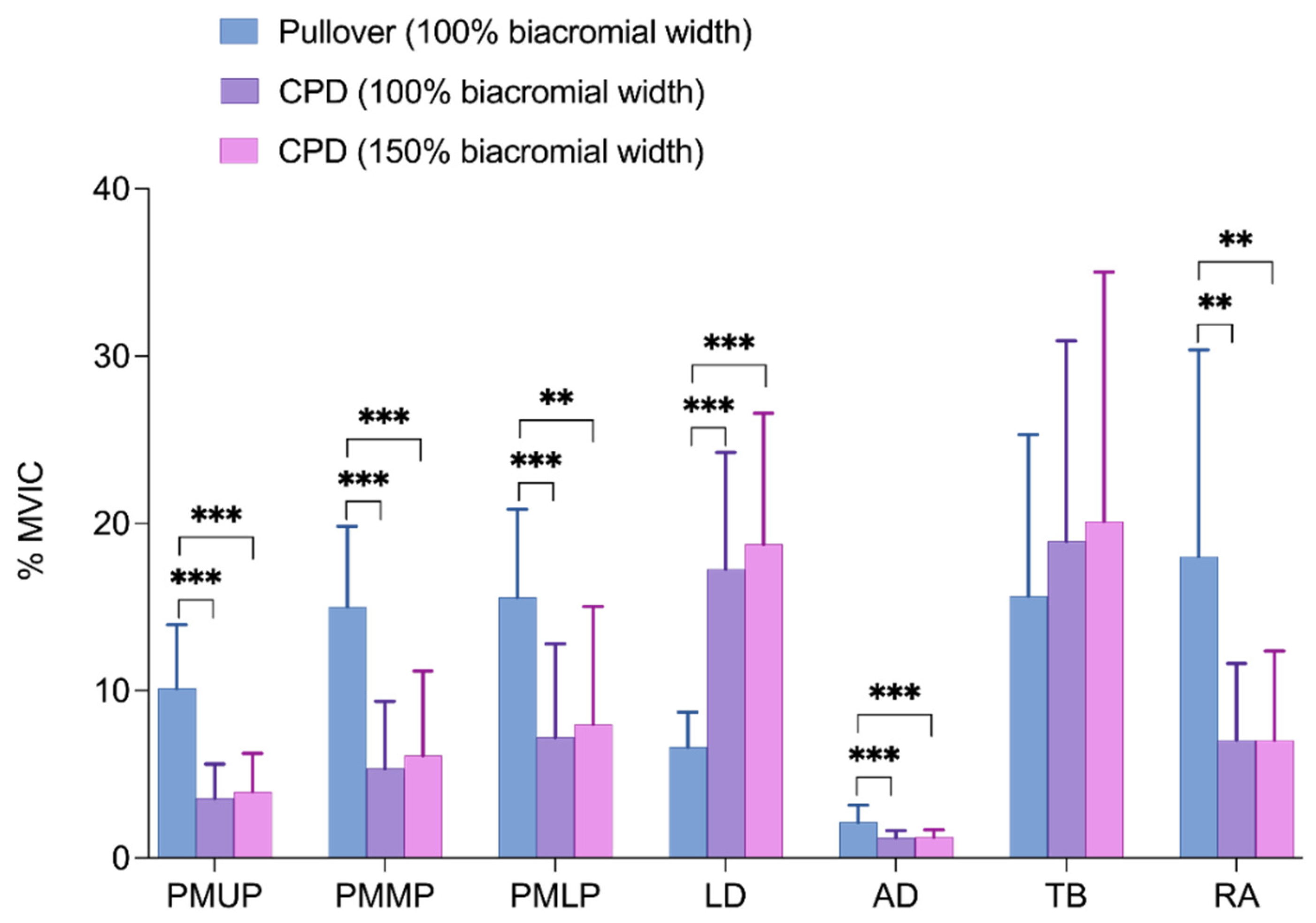
Applied Sciences | Free Full-Text | Comparison of Electromyographic  Activity during Barbell Pullover and Straight Arm Pulldown Exercises
Applied Sciences | Free Full-Text | Comparison of Electromyographic Activity during Barbell Pullover and Straight Arm Pulldown Exercises

Determining the optimal maximal and submaximal voluntary contraction tests  for normalizing the erector spinae muscles
Determining the optimal maximal and submaximal voluntary contraction tests for normalizing the erector spinae muscles
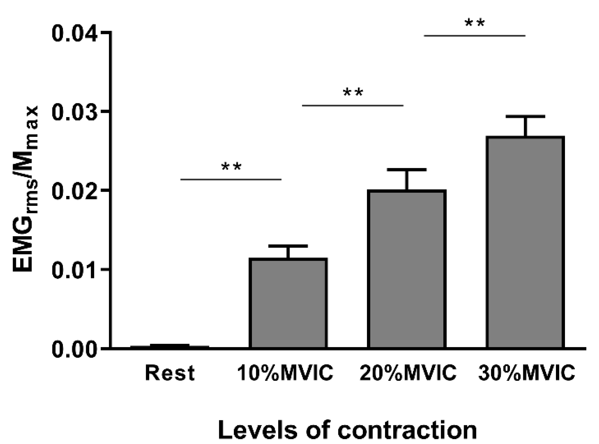
Brain Sciences | Free Full-Text | Influence of Voluntary Contraction Level,  Test Stimulus Intensity and Normalization Procedures on the Evaluation of  Short-Interval Intracortical Inhibition
Brain Sciences | Free Full-Text | Influence of Voluntary Contraction Level, Test Stimulus Intensity and Normalization Procedures on the Evaluation of Short-Interval Intracortical Inhibition
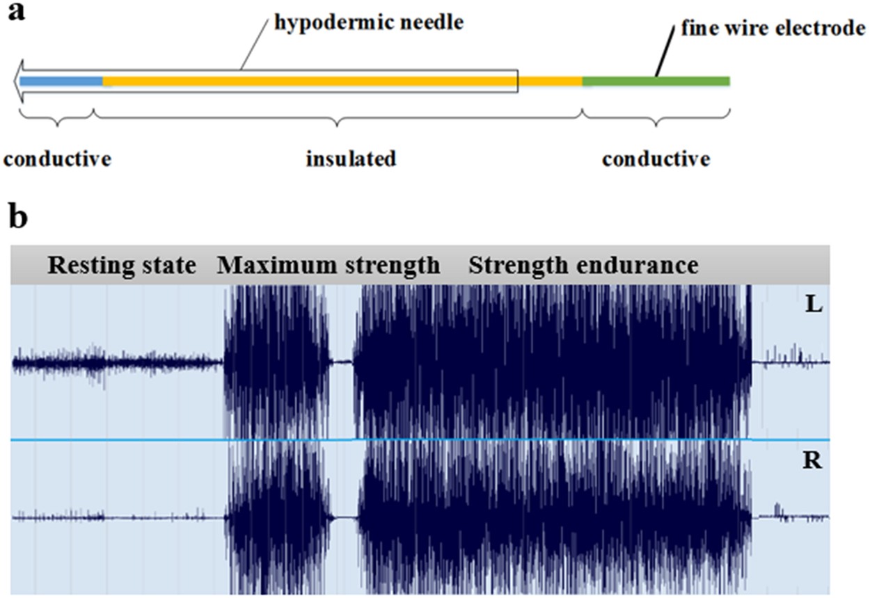
Functional and Morphological Changes in the Deep Lumbar Multifidus Using  Electromyography and Ultrasound | Scientific Reports
Functional and Morphological Changes in the Deep Lumbar Multifidus Using Electromyography and Ultrasound | Scientific Reports

EMG Archives - Steele Lab - University of Washington

Solved 1. Complete the table. Show the equation to calculate | Chegg.com

Percentage of maximum voluntary isometric contraction (%MVIC) of rectus...  | Download Scientific Diagram
Percentage of maximum voluntary isometric contraction (%MVIC) of rectus... | Download Scientific Diagram